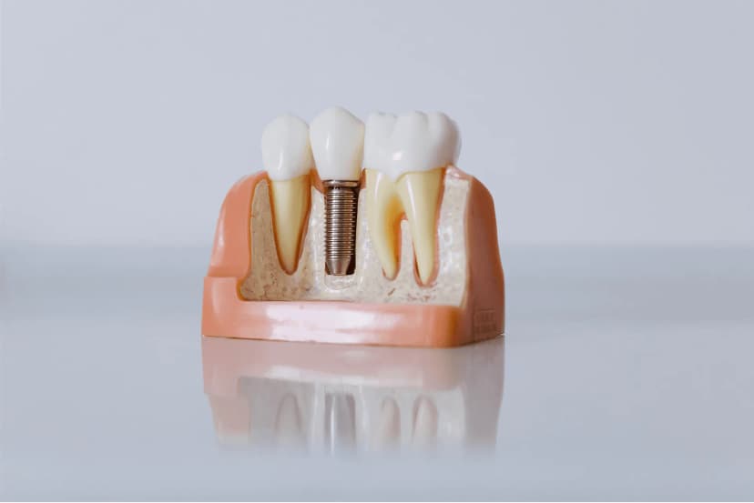
What Is an Impacted Wisdom Tooth?
Impacted wisdom teeth are classified by their position:
- Partially impacted (partial retention): Part of the tooth is visible above the gum but it has not fully erupted. Food debris and infection frequently accumulate beneath the gum flap.
- Fully impacted (total retention): The tooth is completely enclosed in bone or soft tissue and is not visible in the mouth. It is only identified on X-ray.
- Mesioangular (angled forwards): The tooth is angled towards the adjacent second molar. This is the most common impaction pattern and can cause decay or root resorption in the neighbouring tooth.
- Distoangular (angled backwards): The tooth leans towards the back of the jaw. Surgical access can be difficult.
- Horizontal: The tooth lies completely horizontally, pressing firmly on the neighbouring tooth, and usually requires surgical removal.
- Vertical: The tooth is in the correct axis but has not erupted due to lack of space. Extraction is often more straightforward than for other patterns.
Each pattern may call for a different surgical approach. The tooth's position, the shape of its roots and its relationship to neighbouring anatomical structures are assessed in detail using a panoramic X-ray and, where needed, 3-D CBCT imaging.
Wisdom Tooth Pain: Symptoms and What to Do
Wisdom tooth pain arises when an impacted or partly erupted tooth presses on the gum and the adjacent tooth. It is usually felt in the back of the lower jaw and can radiate to the ear and temple.
Home Measures for Wisdom Tooth Pain
- Painkiller: Ibuprofen (400 mg) reduces inflammation and pain
- Salt-water rinse: 3–4 times a day — reduces bacteria under the gum flap
- Cold compress: Apply to the outside of the cheek for 15 minutes
- Mouthwash: A chlorhexidine-based rinse helps control infection
- Soft diet: Avoid hard foods
When to Seek Emergency Care
- Swelling of the face or neck
- Difficulty opening the mouth (trismus)
- Fever (above 38 °C)
- Difficulty swallowing
- Pain that does not respond to painkillers
💡 Important: Even if wisdom tooth pain settles on its own, it is very likely to return. A radiographic assessment should always be made before a decision is taken about extraction.
Recovery After Wisdom Tooth Extraction
First 24 Hours
- Bite on the gauze pack for 30–45 minutes to control bleeding
- Apply cold compresses (20 minutes on, 20 minutes off)
- Eat soft, lukewarm foods
- No smoking, no spitting, no drinking through a straw (dry socket risk)
Days 2–7
- Swelling peaks on days 2–3 and subsides by day 5–7
- Take painkillers regularly
- Rinse gently with salt water from day 2 onwards
- Complete the full course of antibiotics if prescribed
How Long Does Pain Last After Wisdom Tooth Surgery?
| Type of Extraction | Pain Duration | Recovery |
|---|---|---|
| Simple extraction | 2–3 days | 1 week |
| Surgical extraction (partial) | 3–5 days | 2 weeks |
| Surgical extraction (full impaction) | 5–7 days | 2–3 weeks |
Dry Socket (Alveolitis) Risk
A painful complication caused by loss of the blood clot from the socket:
- Symptoms: Severe, throbbing pain intensifying on days 2–4, bad taste/smell
- Risk factors: Smoking, spitting, straw use, oral contraceptives
- Treatment: See your dentist — a medicated dressing is placed in the socket
- Prevention: Avoid smoking and sucking or spitting
Symptoms of an Impacted Wisdom Tooth
Impacted wisdom teeth can sometimes remain silent for years. In many patients, however, the following symptoms develop:
- Pain and throbbing: The commonest symptom — a constant or intermittent ache at the back of the jaw that radiates to the ear or temple, often worse at mealtimes and at night.
- Gum swelling and redness: The soft tissue around a partly erupted tooth becomes inflamed. This condition is called pericoronitis and can progress to serious infection if untreated.
- Bad taste and bad breath: Bacteria and food debris accumulating under the gum flap cause a bad taste and halitosis.
- Difficulty opening the mouth (trismus): Inflammation of the chewing muscles restricts mouth opening, making eating and speaking difficult.
- Sensitivity or decay in the adjacent tooth: The impacted tooth presses on the second molar and can cause root resorption or decay at the contact point.
- Facial swelling: Advanced infection may produce obvious swelling of the lower face, which is a dental emergency.
- Lymph node enlargement: Painful, tender lymph nodes under the jaw may indicate chronic infection.
If you have one or more of these symptoms, we recommend an assessment by an oral surgery specialist. For more information, see our Oral Surgery Guide.
When Is Extraction Needed?
Not every impacted wisdom tooth must be removed. Surgical extraction is indicated in the following situations:
- Recurrent pericoronitis: If you have had two or more infection episodes, antibiotics provide only temporary relief — the definitive treatment is extraction.
- Damage to the adjacent tooth: When the impaction has caused decay, root resorption or a periodontal pocket on the second molar, extraction is planned.
- Cyst or tumour formation: The follicular tissue around the impacted tooth can develop into a dentigerous cyst or ameloblastoma. A follicular space greater than 2.5 mm on imaging is a warning sign.
- Before orthodontic treatment: Impacted wisdom teeth may need to be removed to allow proper tooth alignment, particularly in crowded cases.
- Before prosthodontic or implant treatment: If an implant is planned in that area, or a prosthesis is to be fitted, the impacted tooth must be removed first.
- Unexplained pain: Persistent pain at the back of the jaw with no other cause can be attributed to an impacted tooth.
- No opposing tooth: An erupted wisdom tooth without an opposing tooth supra-erupts and traumatises the opposing gum; it too should be removed.
The decision to extract is made by combining the clinical examination with imaging findings.
Preoperative Assessment
Careful preoperative assessment is essential for safe surgery:
Radiographic Imaging
- Panoramic radiograph (OPG): Shows a general view of all the teeth, the jaws and the surrounding structures. Gives initial information about the tooth's position, number of roots and root morphology.
- 3-D Cone Beam CT (CBCT): Allows millimetric measurement of the distance between the impacted root and the inferior alveolar nerve in the lower jaw. When the root and nerve canal are closely related, 3-D imaging becomes essential.
Proximity to the Nerve Canal
In the lower jaw, the inferior alveolar nerve within the mandibular canal is the most important anatomical risk. This nerve supplies sensation to the lower lip, chin and gum. Where the root superimposes on the nerve canal on the radiograph, the three-dimensional relationship is always assessed with CBCT.
Patient Assessment
- Systemic conditions (diabetes, cardiovascular disease, bleeding disorders)
- Medication (particularly blood thinners and bisphosphonates)
- Allergy history
- Anaesthesia preferences: Most procedures are carried out under local anaesthesia. For patients with dental anxiety, sedation is considered.
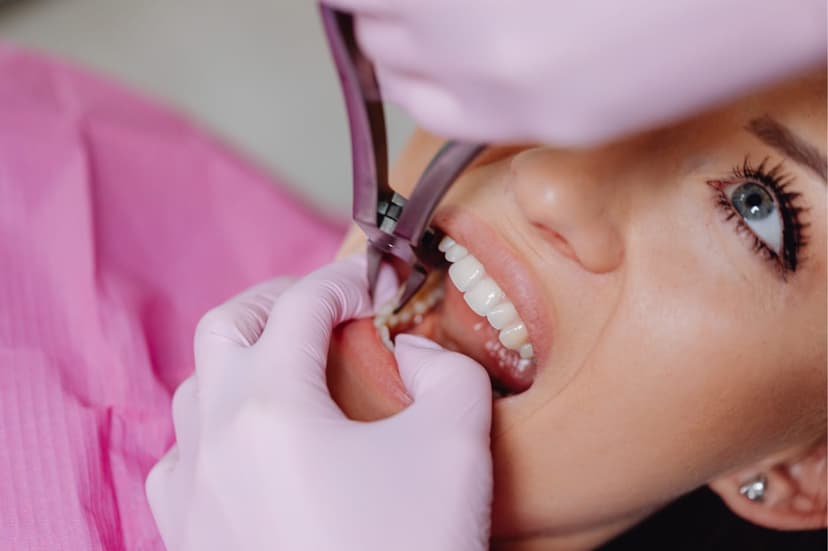
How Is the Surgical Extraction Performed?
Impacted wisdom tooth removal is a controlled surgical procedure consisting of several key steps:
1. Anaesthesia
Local anaesthesia is given to the operative area. An inferior alveolar nerve block is used in the lower jaw and infiltration anaesthesia in the upper jaw. You will feel no pain — only pressure. Conscious sedation or general anaesthesia is available where appropriate.
2. Raising the Flap
A controlled incision is made in the gum and a mucoperiosteal flap is raised. This exposes the bone overlying the impacted tooth. The incision design depends on the tooth's position and the surgeon's experience.
3. Bone Removal (Ostectomy)
The bone covering the tooth is removed in a controlled manner using a surgical bur or piezosurgery (ultrasonic bone cutter). Piezosurgery only cuts hard tissue and does not damage soft tissue, minimising the risk of nerve and vessel injury.
4. Tooth Sectioning (Odontotomy)
Where the tooth cannot be removed in one piece, it is divided into two or more fragments using a bur. This technique is particularly useful for horizontal and mesioangular impactions; it reduces surrounding bone loss and minimises the risk of nerve injury.
5. Removal of the Tooth
The sectioned or loosened tooth is carefully removed from the socket with elevators and forceps.
6. Socket Cleaning and Suturing
The socket is curetted to remove granulation tissue and infected material. A bone graft may be placed where appropriate. The flap is repositioned and closed with resorbable (self-dissolving) sutures which fall out within 7–10 days or are removed at the review appointment.
The whole procedure is usually completed in 20–45 minutes, depending on the degree and pattern of impaction.
Coronectomy as an Alternative
In some cases the roots of the impacted tooth run very close to, or wrap around, the inferior alveolar nerve. Removing the tooth entirely would carry a risk of permanent nerve injury. Coronectomy (intentional partial odontectomy) is a safer alternative for these patients.
In a coronectomy only the crown of the tooth is surgically removed; the roots are left within the bone. Over time the roots migrate deeper into the bone, away from the nerve. If required, the roots can then be removed safely in a second procedure.
Recovery Timeline
Recovery depends on the extent of the surgery and the patient's general health. The following is a general timeline:
Day 1 (Day of Surgery)
- The anaesthesia wears off over 2–4 hours; take care not to bite your lip or tongue during this time.
- Mild bleeding is normal. Bite on the gauze pack for 30–45 minutes.
- Apply ice for the first 24 hours: 15 minutes on, 15 minutes off. This substantially reduces swelling.
- Start the prescribed painkillers and antibiotics on time.
- Eat soft, lukewarm foods. Avoid hot food and drink.
Days 2–3
- Swelling peaks at this stage — this is a normal inflammatory response and begins to settle from day 3.
- Bruising (ecchymosis) may appear, particularly in patients with fair skin, around the cheek and jaw.
- Mouth opening may be restricted; do not force the jaw.
- Continue a soft diet: soup, yoghurt, milk pudding, mashed potato.
First Week
- Pain reduces noticeably. Swelling largely subsides.
- Sutures are checked; if resorbable sutures were used they begin to fall out.
- You can return to light physical activity, but avoid strenuous exercise.
- Whitish granulation tissue forms in the socket; this is not infection but a sign of healing.
Second Week
- Soft-tissue healing is largely complete.
- You can gradually return to a normal diet.
- Mouth opening returns to normal.
- Sutures should have fallen out or been removed.
Complete bone healing takes 6–8 weeks. Following post-operative care instructions substantially speeds recovery.
Possible Complications
As with any surgical procedure, there are some potential complications. When the surgery is performed by an experienced oral surgeon these risks are minimised:
Alveolitis (Dry Socket)
Caused by early disruption or loss of the blood clot in the socket. It presents with severe, throbbing pain on days 3–5. Treatment is placement of a eugenol-based dressing in the socket. Smoking and sucking movements after surgery are the main risk factors.
Bleeding
Mild bleeding in the first 24 hours is normal. Uncontrolled or recurrent bleeding requires medical attention. Patients on blood thinners have their medication adjusted before surgery.
Swelling and Bruising
These are expected responses. However, disproportionate or worsening swelling may indicate infection. If accompanied by fever, urgent assessment is needed.
Nerve Injury (Paraesthesia)
Injury to the inferior alveolar nerve or the lingual nerve during surgery can cause numbness or tingling of the lower lip, chin or tongue. In most cases this is temporary and resolves over weeks to months. Permanent nerve injury is rare (under 1%); preoperative planning with CBCT substantially reduces this risk.
Trismus (Limited Mouth Opening)
Inflammation in the chewing muscles can temporarily restrict mouth opening. Warm compresses and gentle jaw-opening exercises usually resolve this within 1–2 weeks.
Oro-Antral Communication
In upper jaw wisdom tooth extractions the floor of the maxillary sinus can be opened. Small openings close spontaneously; larger ones may require surgical repair.
References
- Sarikov R, Juodzbalys G. Inferior alveolar nerve injury after mandibular third molar extraction: a literature review. J Oral Maxillofac Res. 2014;5(4):e1. PubMed
- He H, Ruan N. Factors influencing inferior alveolar nerve injury after extraction of mandibular third molar. Med Oral Patol Oral Cir Bucal. 2024;29(5):e613-e619. PubMed
- Li Y, et al. Association of the inferior alveolar nerve position and nerve injury: A systematic review and meta-analysis. Healthcare (Basel). 2022;10(9):1782. PubMed
- Rakhshan V. Common risk factors of dry socket following dental extraction: A brief narrative review. J Stomatol Oral Maxillofac Surg. 2018;119(5):407-411. PubMed
- Kuśnierek W, et al. Smoking as a risk factor for dry socket: A systematic review. Dent J. 2022;10(7):121. PubMed
- Cervera-Espert J, et al. Coronectomy of impacted mandibular third molars: A meta-analysis and systematic review. Med Oral Patol Oral Cir Bucal. 2016;21(4):e505-e513. PubMed
- Wehr C, et al. An insight into acute pericoronitis and the need for an evidence-based standard of care. Dent J. 2019;7(3):88. PubMed
Specialist Assessment for Your Impacted Wisdom Tooth
Treatment of an impacted wisdom tooth requires accurate diagnosis and an experienced surgical approach. At Derya Dental Clinic, oral surgery specialist Uzm. Dt. Aykut Gürel carries out a detailed assessment using panoramic X-ray and 3-D CBCT to produce a treatment plan tailored to you.
📞 0216 572 05 20 💬 WhatsApp appointment
Related Guides
For more on oral surgery and tooth extraction:
- Oral Surgery Guide — The full scope of oral and maxillofacial surgery
- Tooth Pain Causes and Treatment — Diagnosis by pain pattern
- What Is Root Canal Treatment — An alternative to extraction
- TMJ (Jaw Joint) Disorders — Jaw joint problems
Related Treatment Pages
This content is for informational purposes only and does not replace medical diagnosis or treatment. Please consult a specialist for decisions about your oral and dental health.