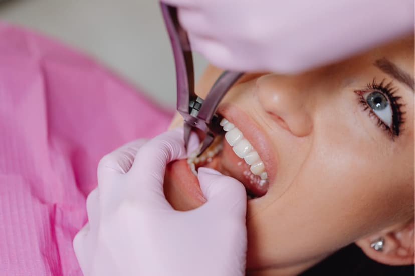
Are Dental X-Rays Safe in Pregnancy?
The American Dental Association (ADA) and the American College of Obstetricians and Gynecologists (ACOG) both state that dental radiography is safe in pregnancy when it is clinically necessary. Modern digital X-ray units deliver very low radiation doses, and lead aprons minimise fetal exposure even further.
Comparative Radiation Doses
| Source | Radiation Dose |
|---|---|
| Digital periapical X-ray | 0.005 mSv |
| Digital panoramic radiograph | 0.01–0.02 mSv |
| Dental CBCT | 0.03–0.20 mSv |
| Daily natural background radiation | 0.008 mSv |
| Air travel (Istanbul–London) | 0.02 mSv |
As the table shows, the dose from a single digital periapical X-ray is lower than a single day's natural background radiation.
When Is a Dental X-Ray Necessary in Pregnancy?
During pregnancy, dental X-rays should only be taken when there is a clinical need:
- Dental emergency — suspected abscess or advanced infection
- Trauma — tooth fracture or jaw injury
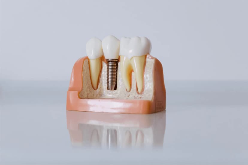
- Need for root canal treatment — imaging is essential for planning
- Surgical assessment — suspected impacted tooth or cyst
When Can an X-Ray Be Postponed?
- Routine review radiographs (6-monthly periodic imaging)
- Imaging for cosmetic treatment planning
- Non-urgent implant planning
- Extensive CBCT — where possible, deferred until after delivery
Which Trimester Is Safest for X-Rays?
| Trimester | Period | Approach |
|---|---|---|
| First trimester (0–12 weeks) | Organogenesis | Emergencies only; otherwise postpone where possible |
| Second trimester (13–27 weeks) | Safest window | Necessary treatment and imaging are planned for this period |
| Third trimester (28–40 weeks) | Late pregnancy | Short procedures are fine; longer work is deferred until after delivery |
The second trimester is the ideal window. Organogenesis is complete, and the uterus has not yet grown to the size where extended supine positioning is uncomfortable.
Protective Measures
The following measures are standard whenever a dental X-ray is taken during pregnancy:
- Lead apron — a lead apron covering the abdomen and pelvis is always used
- Thyroid collar — a thyroid shield is placed around the neck
- Digital sensors — 70–80% lower dose than conventional film
- Minimum exposures — only the clinically necessary regions are imaged
- Short exposure time — kept to the minimum required
Why Oral Health Matters in Pregnancy
Hormonal changes during pregnancy increase the risk of:
- Gingivitis (pregnancy gingivitis)
- Accelerated tooth decay (nausea, vomiting, acidic snacking)
- Pregnancy epulis (pyogenic granuloma)
- Periodontal disease, which has been linked with preterm birth and low birth weight
For these reasons, postponing dental care in pregnancy can be riskier than providing it. An untreated abscess puts both mother and baby at risk.
Conclusion
Dental X-rays during pregnancy can be taken safely when they are clinically justified and the proper precautions are in place. The key is not to postpone necessary treatment unnecessarily, and to make the decision based on clinical assessment. For any oral health problems during pregnancy, please contact our clinic.
This article was written by Dr Aykut Gürel. It is intended for information only and does not replace individual medical advice.
Related Treatment Pages
This content is for informational purposes only and does not replace medical diagnosis or treatment. Please consult a specialist for decisions about your oral and dental health.